


Benign growths can develop in many tissues, but not all lumps are created equal. Uterine fibroids (leiomyomas) and breast fibroadenomas are both non-cancerous, yet their origins, symptoms, and management differ significantly. Understanding these distinctions is vital for proper diagnosis and treatment.
1. Tissue Origin & Pathophysiology

- Fibroids: Arise from smooth muscle cells in the uterine wall, growing under estrogen and progesterone influence .
- Fibroadenomas: Originate from glandular and stromal breast tissue, often linked to hormonal fluctuations in younger women .
2. Clinical Presentation & Symptoms

|
Feature |
Fibroids |
Fibroadenomas |
|---|---|---|
|
Age Group |
30 - 50 years |
15 - 35 years |
|
Pain & Discomfort |
Heavy bleeding, cramps, bloating |
Usually painless, mobile lump |
|
Palpable Lump |
Rarely felt unless large |
Easily felt, firm, rubbery |
|
Systemic Effects |
Anemia, fatigue |
Minimal |
3. Diagnostic Techniques

- Ultrasound (US): First-line for fibroids; shows size and location within uterus .
- Mammography & US: Standard for breast lumps; fibroadenomas appear as well-circumscribed, homogenous nodules .
- MRI & Biopsy: Used when imaging is inconclusive or tissue sampling is needed.
4. Treatment Approaches

- Fibroids: Watchful waiting, GnRH analogs, Uterine Fibroid Embolization (UFE), myomectomy, or hysterectomy .
- Fibroadenomas: Often monitored; can be removed via excisional biopsy or cryoablation if symptomatic or growing .
5. When to Seek Care
- Fibroids: Heavy bleeding, severe pain, fertility issues → gynecologist evaluation.
- Fibroadenomas: New breast lump, rapid growth, or family history of breast cancer → breast specialist.
Next Steps:
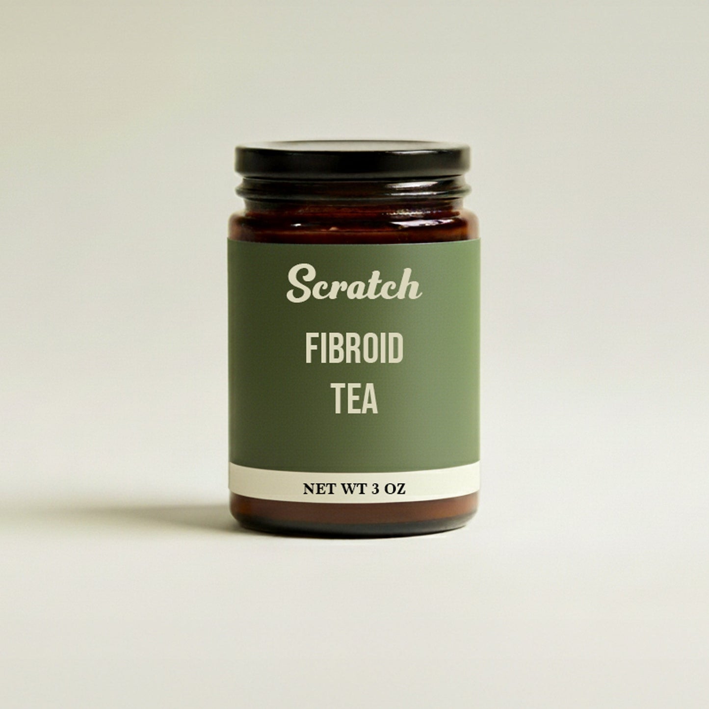
For a step-by-step integrative approach including natural, plant-based protocols try our Fibroid Wellness Collection Bundle .
References
- NIH: Uterine Fibroids Fact Sheet
- National Library of Medicine: Fibroadenoma Overview
- CDC: Fibroid Data & Statistics
- FDA: Imaging Fibroids
- American Cancer Society: Breast Imaging Guidelines
- Mayo Clinic: Fibroadenoma Treatment